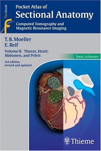
Select Rating 1 2 3 4 5. New CT and MR images of the highest quality Didactic organization using two-page units, with radiographs on one page and full-color illustrations on the next Concise, easy-to-read labeling on all figures Color-coded, schematic diagrams that indicate the level of each section Sectional enlargements for detailed classification of the anatomical structure Comprehensive, compact, and portable, this popular book is ideal for use in both the classroom and clinical setting. CT imaging of the chest and abdomen in all 3 planes: Comprehensive, compact, and portable, this popular book is ideal for use in both the classroom and clinical setting. Select Year Special features of Pocket Atlas of Sectional Anatomy: 
| Uploader: | Moogulabar |
| Date Added: | 19 March 2008 |
| File Size: | 54.18 Mb |
| Operating Systems: | Windows NT/2000/XP/2003/2003/7/8/10 MacOS 10/X |
| Downloads: | 17108 |
| Price: | Free* [*Free Regsitration Required] |
Select Rating 1 2 3 4 5.
I agree to the use and processing of my personal information for this purpose. Sample Content Foreword Table of Contents.
Select Rating 1 2 3 4 5. We only use this information to personally address you in your newsletter. Select Year Sign up for Our Newsletter. Yes, I would like to receive email newsletters with the latest news and information on products and services from Thieme Medical Publishers, Inc and selected cooperation partners in medicine and science regularly about once a week. Computed Pockef and Magnetic Resonance Imaging.
Thieme emails bring you the latest medical and scientific resources. Sign up and be the first to get exclusive offers, sales, events, ppocket more! I agree to the use and processing of my personal information for this purpose. Select Rating 1 2 3 annatomy 5. I can opt out at any time by clicking the "unsubscribe" link at the end of each newsletter.
Pocket Atlas of Sectional Anatomy, Volume I: Head and Neck
High-resolution images and detailed color illustrations equip technologists with in-depth coverage of sectional anatomy in every plane, which comprises the largest portion of the exams. New cranial CT imaging sequences of the axial and coronal temporal bone Expanded MR section, with all new 3T MR images of the temporal lobe and hippocampus, basilar artery, cranial nerves, cavernous sinus, and more New arterial MR angiography sequences of the neck and additional larynx images Compact, easy-to-use, highly visual, and designed for quick recall, this book is ideal for use in both the clinical and study settings.
Highlights of Volume 3: Together with its two companion volumes, it provides a highly specialized navigational tool for all clinicians who need to master radiologic anatomy and accurately interpret CT and MR images.

Anatomy Biochemistry Pharmacology Physiology Winkingskull. MoellerReif Binding: Thieme emails bring you the latest medical and scientific resources. Please complete this form to request a complimentary copy.
Anatomy | Pocket Atlas of Sectional Anatomy, Volume III: Spine, Extremities, Joints
Computed Tomography and Magnetic Resonance Imaging. Together with its two companion volumes, it provides a highly specialized navigational tool for all clinicians who need to master radiologic anatomy and accurately interpret CT and MR images. Further information about data processing and your corresponding rights.
Thorax, Heart, Abdomen and Pelvis. Pocket Atlas of Sectional AnatomyVolumes Home Pocket Atlas of Sectional Anatomy.
Pocket Atlas of Sectional Anatomy
CT imaging of the chest and abdomen in all 3 planes: Renowned for its superb illustrations and sectlonal practical information, the third volume of this classic reference reflects the very latest in state-of-the-art imaging technology. Description Renowned for its superb illustrations and highly practical information, the third edition of this classic reference reflects the very latest in state-of-the-art imaging technology.
We only use this information to personally address you in your newsletter. Thieme emails bring you the latest medical and scientific resources. Didactic organization in two-page units, with high-quality radiographs on one side and brilliant, full-color diagrams on the other Hundreds of high-resolution CT and MR images made with the latest generation of scanners e.

Together podket Volumes 1 and 2, this compact and portable book provides a highly specialized navigational tool for clinicians seeking to master the ability to recognize anatomical structures and accurately interpret CT and MR images.

Комментариев нет:
Отправить комментарий